Animal Cell Electron Microscope / Pin em PinBio_1002.2016 / Recent experimentation has been aimed at utilizing animal cells.
byLeon Avarbuch-0
Animal Cell Electron Microscope / Pin em PinBio_1002.2016 / Recent experimentation has been aimed at utilizing animal cells.. Light and electron microscopes allow us to see inside cells. However, they usually can achieve a maximum of 2000x magnification which is not sufficient to see many other tiny organelles. Generalized cell is used for structure of animal cell and plant cell. Ultrastructure is the architecture of cells that is visible at higher magnifications than found on a standard light microscope. A cell is a very tiny structure which exists in living bodies.
Scanning electron microscope cell images. Light and electron microscopes allow us to see inside cells. Animal, plant and bacterial cells. Ishita observed a slide of eukaryotic cell under electron microscope. The ultrastructure of cells viewed by transmission electron microscopy and scanning electron microscopy.
Labeled Plant Cell Under Electron Microscope from www.vcbio.science.ru.nl .cells (seen through a scanning electron microscope) are from very different organisms, yet all share certain characteristics of basic cell structure. Bring your presentation to life. Animal cells are of various sizes and have irregular shapes. However, they usually can achieve a maximum of 2000x magnification which is not sufficient to see many other tiny organelles. How is it different from animal cell? Image:animal cell seen under electron microscope. There are many types of cells, all grouped into one of two broad categories: 1st john 1:1 holy hydrogen light of creation has been discovered glowing within the human cell wall plasma nucleus as seen with an electron microscope in.
Animal cells are of various sizes and have irregular shapes.
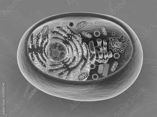
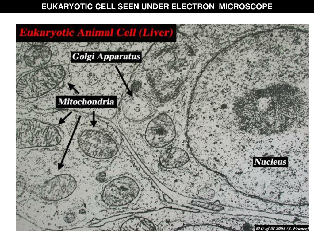
Ishita observed a slide of eukaryotic cell under electron microscope. Although the very first electron microscopy (em) images of eukaryotic cells were attributed in 1945, it was the ruska family that not only developed the em, but also pioneered in the field of infections with pictures of bacteria and viruses. Here is an electron micrograph of an animal cell with the labels superimposed: An electron microscope is a microscope that uses a beam of accelerated electrons as a source of illumination. Methylene blue is often used for animal cells there are two kinds of electron microscope. Light and electron microscopes allow us to see inside cells. Image:animal cell seen under electron microscope. 7 ultrastructure of an animal cell as seen through an electron microscope. Resolving power is the ability to distinguish between separate things which are close to each other. The transmission electron microscope (tem) works. The ultrastructure of cells viewed by transmission electron microscopy and scanning electron microscopy. Recent experimentation has been aimed at utilizing animal cells. Animal, plant and bacterial cells.
However, they usually can achieve a maximum of 2000x magnification which is not sufficient to see many other tiny organelles. Image:animal cell seen under electron microscope. Resolving power is the ability to distinguish between separate things which are close to each other. Animal, plant and bacterial cells. Image:plant cell seen under electron microscope.
Animal cell, 3d rendering, Scanning Electron Microscope ... from as2.ftcdn.net Anatomy_and_physiology_of_animals_animal_cell_electron_microscope.jpg (557 × 540 pixels, file size: .cells (seen through a scanning electron microscope) are from very different organisms, yet all share certain characteristics of basic cell structure. You see that many features are in common. Animal cell (as seen under electron microscope). However, light microscopes form real colour images and can be used to watch living processes occur in microscopic detail, while electron microscopes cannot be used to study living. Here is an electron micrograph of an animal cell with the labels superimposed: A generalised animal cell as observed under an electron microscope. Light and electron microscopes allow us to see inside cells.
You see that many features are in common.
Image:animal cell seen under electron microscope. Ishita observed a slide of eukaryotic cell under electron microscope. A generalised animal cell as observed under an electron microscope. However, light microscopes form real colour images and can be used to watch living processes occur in microscopic detail, while electron microscopes cannot be used to study living. There are many types of cells, all grouped into one of two broad categories: Although the very first electron microscopy (em) images of eukaryotic cells were attributed in 1945, it was the ruska family that not only developed the em, but also pioneered in the field of infections with pictures of bacteria and viruses. The cell theory, or cell doctrine, states that all organisms are composed of. There is also another type of microscope called light microscope which uses light. Ultrastructure is the architecture of cells that is visible at higher magnifications than found on a standard light microscope. Generalized cell is used for structure of animal cell and plant cell. Cell cycle math enrichment transition words worksheet busy teacher possessive nouns worksheets measurement worksheets decimals worksheets kids worksheets printables worksheets. Light and electron microscopes allow us to see inside cells. Methylene blue is often used for animal cells there are two kinds of electron microscope.
There are two types of electron microscope Of all the techniques used in biology microscopy is probably the most different stains are used for different types of tissues. Animal cell (as seen under electron microscope). The ultrastructure of cells viewed by transmission electron microscopy and scanning electron microscopy. Red blood cells under 100x and 400x microscope.
Electron Microscope Eukaryotic Animal Cell - Micropedia from image2.slideserve.com The animal cell is more. 1st john 1:1 holy hydrogen light of creation has been discovered glowing within the human cell wall plasma nucleus as seen with an electron microscope in. There are many types of cells, all grouped into one of two broad categories: Some disadvantage of electron microscopes are that they cannot display living specimens in natural colours. A generalised animal cell as observed under an electron microscope. An electron microscope is a microscope that uses a beam of accelerated electrons as a source of illumination. The animal cell is more with a transmission electron microscope (tem) and generic contrast staining (osmium, uranyl, lead) of a section through a cell you will not only see. Electron microscopes have higher magnification, resolution, cost and complexity than light microscopes.
Resolving power is the ability to distinguish between separate things which are close to each other.
Animal, plant and bacterial cells. Image:animal cell seen under electron microscope. Anatomy_and_physiology_of_animals_animal_cell_electron_microscope.jpg (557 × 540 pixels, file size: Electron microscopes use electron beams focused by electromagnets to magnify and resolve microscopic specimens. Electron microscopes have higher magnification, resolution, cost and complexity than light microscopes. However, light microscopes form real colour images and can be used to watch living processes occur in microscopic detail, while electron microscopes cannot be used to study living. Although the very first electron microscopy (em) images of eukaryotic cells were attributed in 1945, it was the ruska family that not only developed the em, but also pioneered in the field of infections with pictures of bacteria and viruses. Generalized cell is used for structure of animal cell and plant cell. The animal cell is more. Scanning electron microscope cell images. Electron microscope uses electrons and an ordinary microscope uses simple glass plate. Typical animal cell pinocytotic vesicle lysosome golgi vesicles golgi vesicles rough er (endoplasmic reticulum) smooth er (no ribosomes) cell (plasma) membrane… download this awesome diagram. Resolving power is the ability to distinguish between separate things which are close to each other.


Post a Comment